
Welcome to Wellspring Health and Wellness’s patient resource on ankle problems.
The ankle joint functions like a hinge, but it is much more complex. It consists of several important structures that make it very stable. This stability is crucial as the ankle must support 1.5 times your body weight when you walk and up to eight times your body weight when you run.
Normal ankle function is essential for a smooth and effortless gait. The muscles, tendons, and ligaments that support the ankle joint work together to propel the body. Any condition that disrupts the normal functioning of the ankle can make daily activities difficult and painful.
This guide will help you understand:
- The parts that make up the ankle
- How the ankle works
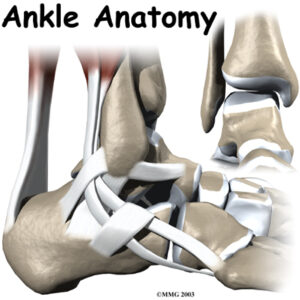
Important Structures
The important structures of the ankle can be divided into several categories, including:
- Bones and joints
- Ligaments and tendons
- Muscles
- Nerves
- Blood vessels
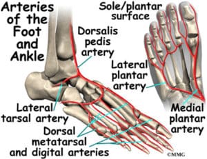
The top of the foot is referred to as the dorsal surface, while the sole of the foot is the plantar surface.
Ankle Bones
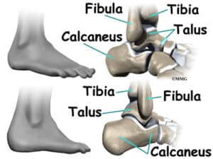
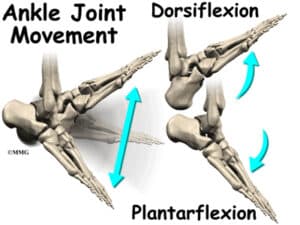
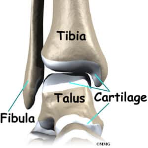
The ankle joint is formed by the connection of three bones. The ankle bone called the talus, fits into a socket formed by the lower end of the tibia (shinbone) and the fibula (the small bone of the lower leg). The bottom of the talus sits on the heel bone, called the calcaneus. The talus acts like a hinge within this socket, allowing the foot to move up (dorsiflexion) and down (plantarflexion).
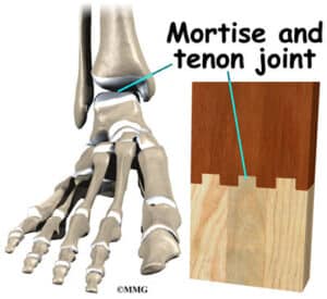
Talus Works Like a Hinge
Woodworkers and craftsmen use similar construction methods, called mortise and tenon, to create stable structures. This design is used to make strong and sturdy items, such as furniture and buildings.
Inside the joint, the bones are covered with articular cartilage, a slick material that allows the bones to move smoothly against one another. This cartilage is about one-quarter of an inch thick in most weight-bearing joints, like the ankle, hip, or knee. It is soft enough for shock absorption but tough enough to last a lifetime unless injured.
Ligaments of the Ankle
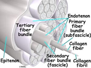
Ligaments are soft tissues that attach bones to bones, similar to tendons, which attach muscles to bones. Both are made up of small fibers of collagen bundled together to form a rope-like structure. The thickness of the ligament or tendon determines its strength.
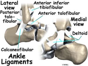
Ligaments on both sides of the ankle joint help hold the bones together. Three ligaments make up the lateral ligament complex on the side of the ankle, which is farthest from the other ankle. These include the anterior talofibular ligament (ATFL), the calcaneofibular ligament (CFL), and the posterior talofibular ligament (PTFL). The medial side (closest to the other ankle) is supported by a thick ligament called the deltoid ligament.
Ligaments also support the lower end of the leg, which forms a hinge for the ankle. This series of ligaments support the ankle syndesmosis, the part where the fibula meets the tibia. Three main ligaments support this area: the anterior inferior tibiofibular ligament (AITFL), the posterior inferior tibiofibular ligament (PITFL), and the transverse ligament. The interosseous ligament, a long sheet of connective tissue, lies between the tibia and fibula, connecting them from the knee to the ankle.

The ligaments around the ankle joint help form the joint capsule, a watertight sac made up of ligaments and soft tissues that fill in the gaps and form the sac.
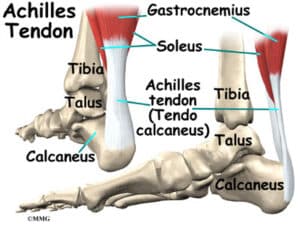
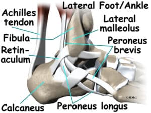
The ankle joint is also supported by tendons. The large Achilles tendon, which attaches the calf muscles to the calcaneus (heel bone), is crucial for walking, running, and jumping. The posterior tibial tendon, attaching one of the smaller calf muscles to the underside of the foot, supports the arch and allows us to turn the foot inward.
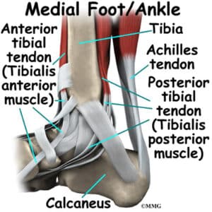
The anterior tibial tendon allows us to raise the foot, and two tendons called the peroneals run behind the outer bump of the ankle, helping to turn the foot down and out.
Muscles of the Ankle
Most of the ankle’s motion is caused by the stronger muscles in the lower leg, whose tendons pass by the ankle and connect in the foot. Contraction of these muscles is the primary way we move our ankles when we walk, run, and jump.
The key ankle muscles and their actions include:
- The peroneals (peroneus longus and peroneus brevis) are on the outside edge of the ankle and foot, which bend the ankle down and out.
- The calf muscles (gastrocnemius and soleus) connect to the calcaneus by the Achilles tendon and bend the ankle down when tightened.
- The posterior tibialis muscle supports the arch and helps turn the foot inward.
- The anterior tibialis pulls the ankle upward.
Nerves of the Ankle
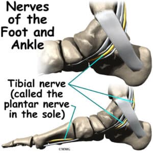
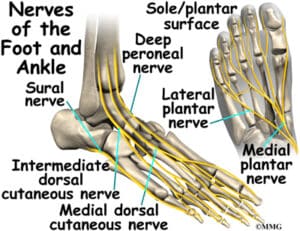
The nerve supply of the ankle comes from nerves that pass by the ankle on their way into the foot. The tibial nerve runs behind the medial malleolus. Another nerve crosses in front of the ankle on its way to the top of the foot. There is also a nerve that passes along the outer edge of the ankle. These nerves control the muscles in this area and provide sensation to the top and outside edge of the foot.
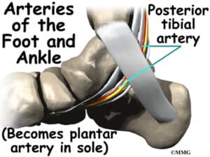
Blood Vessels of the Ankle
The ankle receives blood from nearby arteries that pass by the ankle on their way to the foot. The dorsalis pedis runs in front of the ankle to the top of the foot, where you can feel your pulse. Another large artery, called the posterior tibial artery, runs behind the medial malleolus and supplies smaller blood vessels to the inside edge of the ankle joint. Other less important arteries also supply blood to the ankle.
As you can see, the anatomy of the ankle is very complex. When everything works together, the ankle functions correctly. When one part becomes damaged, it can affect every other part of the ankle and foot, leading to problems.